
Shoulder Dislocation vs Shoulder Fracture: How to Tell the Difference
Shoulder Dislocation vs Shoulder Fracture: How to Tell the Difference
A heavy fall, a sports collision, a road accident — and suddenly your shoulder is in agony. You can't move your arm. Something is visibly wrong. But is the bone broken, or has the joint come out of place? The answer matters enormously, because a dislocation and a fracture are different injuries that require different treatments — and confusing the two, or attempting the wrong first-aid response, can make things significantly worse.
Here is a clear, medically grounded guide to understanding the difference between a shoulder dislocation and a shoulder fracture — their causes, symptoms, how doctors tell them apart, and what treatment involves.
Understanding the Anatomy First
The shoulder is the most mobile joint in the human body, which is precisely why it is also one of the most injury-prone. It consists of three bones: the clavicle (collarbone), the scapula (shoulder blade), and the proximal humerus — the rounded upper part of the arm bone. These bones intersect to form two joints in the shoulder.
The key joint is the glenohumeral joint — a ball-and-socket arrangement where the rounded head of the humerus sits within a shallow cup (the glenoid) on the scapula. Surrounding this joint are the rotator cuff muscles, ligaments, and the labrum — a cartilage ring that deepens the socket and adds stability.
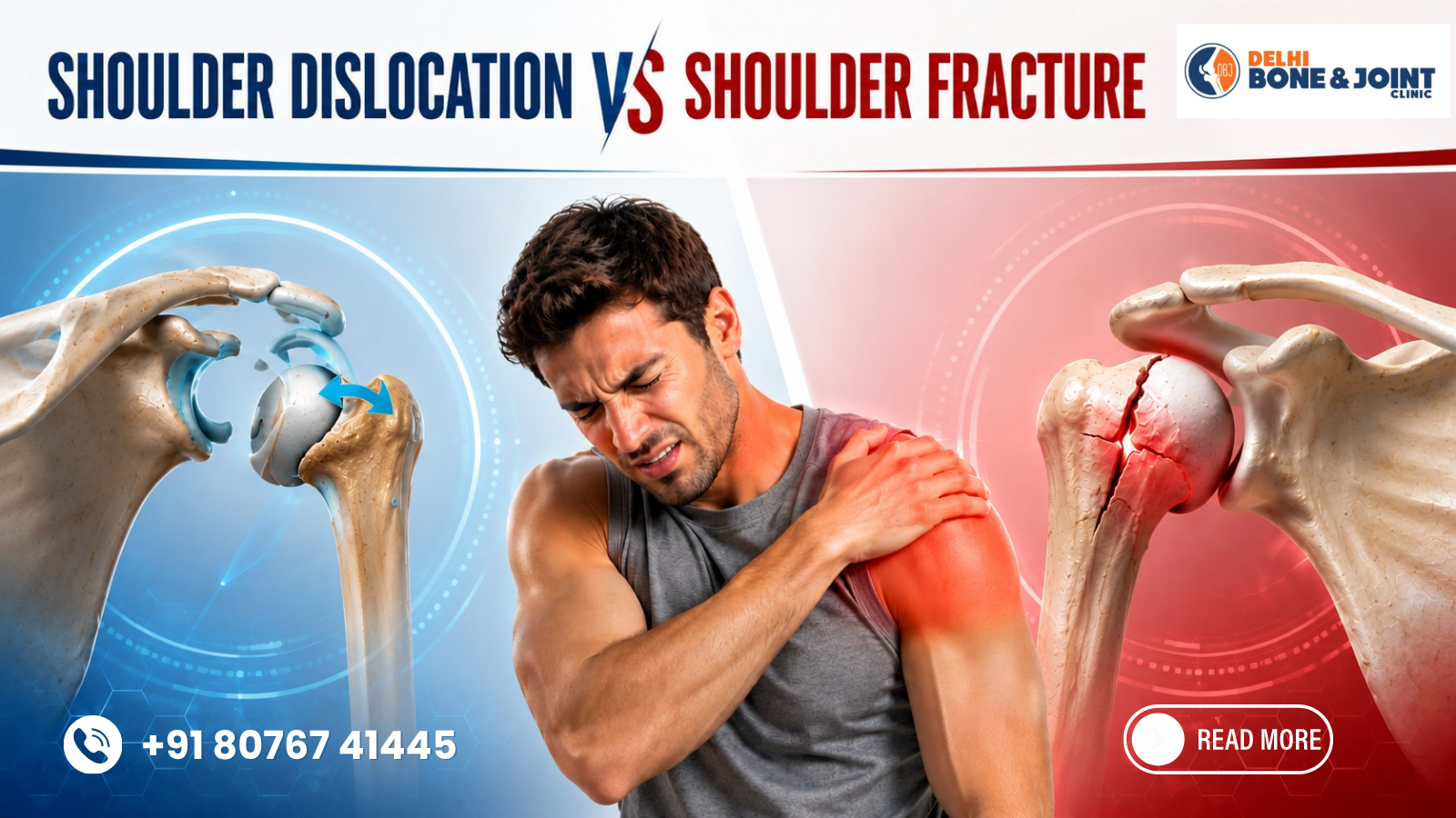
A fracture is a break in one or more of the bones. A dislocation is when the ball of the humerus slips out of the socket entirely. These are distinct injuries — though they can occasionally occur simultaneously, making accurate diagnosis even more critical.
What Is a Shoulder Dislocation?
Traumatic anterior shoulder dislocation is the most common type of joint dislocation, with an incidence of 11 to 29 per 100,000 persons per year. Shoulder dislocations are anterior in 95% or more of patients; the mechanism is abduction and external rotation. In practical terms, this typically happens when the arm is forced outward and upward — as in breaking a fall with an outstretched arm, taking a tackle in sport, or suffering a blow to the back of the shoulder.
In an anterior dislocation, the humeral head slips forward and downward out of the glenoid socket. Posterior dislocations — where the head slips backward — are far less common and are more often associated with seizures or electric shocks.
Key Symptoms of a Shoulder Dislocation
Visible deformity: The shoulder contour changes dramatically. The normal rounded shape of the shoulder is lost; the acromion becomes prominent, and the humeral head cannot be palpated in its usual position. There may be a hollow or "squaring off" of the shoulder profile.
Immediate, severe pain: Onset is instantaneous at the moment of injury, often accompanied by a popping or tearing sensation.
Arm held in a fixed position: Patients are unwilling to move the arm. The elbow is typically held slightly away from the body in partial abduction.
Muscle spasm: The surrounding muscles go into intense spasm, attempting to protect the joint, adding to the pain and resistance to movement.
Numbness or tingling: The axillary nerve runs close to the shoulder joint and is vulnerable to stretch injury during dislocation. Numbness over the outer upper arm (the "regimental badge" area) is a classic sign of axillary nerve involvement and must be checked before any reduction attempt.
What Is a Shoulder Fracture?
A shoulder fracture is a break in any of the three bones that make up the shoulder complex. Fractures commonly involve the clavicle (collarbone), the proximal humerus (top of the upper arm bone), and the scapula (shoulder blade).
Clavicle fractures are the most common shoulder fracture, particularly in children, adolescents, and cyclists. They result from a direct blow to the shoulder or a fall onto an outstretched hand. People with a clavicle fracture usually find it difficult or painful to raise the arm, and bruising, swelling, or a visible bump may appear along the collarbone.
Proximal humerus fractures — breaks at the top of the upper arm bone — are especially common in adults over 65, where osteoporosis lowers the bone's resistance to impact. Proximal humerus fractures are more common in people older than 65 or those who have bone density issues. A relatively minor fall that would produce only a bruise in a younger person can cause a complex multi-part fracture in an older adult.
Scapula fractures are rare because the shoulder blade is so well protected by surrounding musculature. Scapula fractures usually result from a bad collision in a contact sport or automobile accident, and patients who sustain this type of fracture are more likely to also have nerve injuries, rib fractures, or lung injuries.
Key Symptoms of a Shoulder Fracture
Localised, point-specific tenderness: Unlike a dislocation, where pain is diffuse across the joint, fracture pain is often most intense at one specific bony point — along the clavicle, at the top of the humerus, or across the shoulder blade.
Swelling and bruising: Develops rapidly over the fracture site and may spread across the upper arm, chest wall, or shoulder region over 24–48 hours.
Crepitus: A grating or crunching sensation when the shoulder region is gently palpated — caused by broken bone ends moving against each other. This should never be deliberately tested.
Inability to move the arm: Present in both fractures and dislocations, but with a fracture, even passive movement (someone else moving the arm gently) causes severe pain, whereas in a dislocation, passive movement may be slightly less restricted once muscle spasm subsides.
Visible bump or deformity along the bone: A clavicle fracture often produces a visible bump or lump along the collarbone rather than the hollow contour deformity of a dislocation.
The Critical Diagnostic Overlap — and Why X-Ray Is Essential
Here is the fundamental clinical challenge: shoulder dislocations and fractures share several major symptoms — severe pain, swelling, bruising, and inability to move the arm. Even experienced clinicians cannot always distinguish between them reliably on physical examination alone.
Fractures of the clavicle or proximal humerus can be caused by a direct blow from a fall, collision, or motor vehicle accident — the same mechanisms that cause dislocations. And critically, the two injuries frequently co-exist: a dislocation can cause an avulsion fracture of the greater tuberosity (a piece of bone pulled off during dislocation), and a proximal humerus fracture can occur simultaneously with glenohumeral dislocation.
This co-existence is precisely why attempting to "pop a dislocated shoulder back" without X-ray confirmation first is dangerous. If a fracture is also present, attempting reduction can displace bone fragments, damage blood vessels, or worsen nerve injury.
An X-ray is always the mandatory first step. It confirms dislocation direction, identifies fractures, and determines whether a fracture-dislocation is present. A CT scan provides additional detail for complex fractures — assessing the number of fragments, displacement, and joint involvement. MRI is reserved for soft tissue assessment, including rotator cuff tears and labral injuries that frequently accompany both injury types.
Treatment: How They Differ
Treating a Dislocation
The primary treatment for a shoulder dislocation is closed reduction — a technique where the humeral head is guided back into the glenoid socket under sedation or regional anaesthesia. This is performed by a trained clinician using one of several established techniques. Once reduced, the shoulder is immobilised in a sling for several weeks, followed by a structured physiotherapy programme to restore strength and stability. Young patients with first-time dislocations face a high recurrence rate; those with repeated instability may be candidates for arthroscopic stabilisation surgery (Bankart repair).
Treating a Fracture
Fracture management depends on fracture type, severity, displacement, and the patient's age and bone quality. Many clavicle fractures and minimally displaced proximal humerus fractures are managed conservatively — with a sling, pain management, and progressive physiotherapy. Displaced or multi-fragment fractures typically require surgical fixation using plates, screws, or intramedullary nails. Complex proximal humerus fractures in elderly patients with poor bone quality may be best treated with shoulder replacement (hemiarthroplasty or reverse total shoulder arthroplasty).
For patients in West Delhi seeking the best shoulder fracture treatment in Janakpuri, early evaluation at a facility with orthopaedic imaging and surgical capability ensures that the correct treatment pathway — conservative or surgical — is identified before any window for optimal outcome is missed.
When to Seek Emergency Care
Both injuries require prompt medical attention, but certain features demand urgent or emergency evaluation:
Any visible deformity of the shoulder or upper arm
Numbness, tingling, or weakness in the hand or arm (possible nerve or vascular injury)
Pale, cold, or pulseless hand after shoulder injury (arterial injury — surgical emergency)
Open fracture where bone is visible through the skin
High-energy mechanism (road accident, significant fall from height)
Do not attempt to reduce a dislocated shoulder yourself or allow an untrained person to do so. Do not apply tight bandages that could impair circulation. Support the arm in the position of comfort with a soft sling and proceed to an emergency department or orthopaedic specialist immediately.
Rehabilitation: The Overlooked Phase
Whether the final diagnosis is a dislocation or fracture, rehabilitation is not optional — it is essential. Shoulder injuries that are adequately treated surgically but poorly rehabilitated frequently result in stiffness, weakness, chronic instability, and long-term functional limitation.
A structured physiotherapy programme — progressing from early pendulum exercises to rotator cuff strengthening to sport-specific or activity-specific training — is the bridge between treatment and full recovery. Recovery timelines vary: clavicle fractures typically heal in 6–12 weeks; proximal humerus fractures may take 3–6 months; surgically repaired dislocations often require 4–6 months before return to contact sport is safe.
The best shoulder fracture treatment in Janakpuri integrates orthopaedic surgical expertise with structured post-operative rehabilitation — because the quality of long-term recovery depends as much on what happens after treatment as on the procedure itself.
FAQs
Q1. Can you move your arm with a dislocated shoulder but not a fracture?
Not reliably — both injuries severely restrict arm movement. The key difference is visible joint deformity in dislocation versus point tenderness over a specific bone in fracture, but an X-ray is always needed to confirm.
Q2. Can a shoulder dislocation and fracture happen at the same time?
Yes — fracture-dislocations are relatively common, particularly avulsion of the greater tuberosity during anterior dislocation. This is why X-ray before reduction is non-negotiable.
Q3. How long does a dislocated shoulder take to heal vs a fracture?
Simple dislocations typically resolve in 6–12 weeks with physiotherapy. Fractures range from 6 weeks (undisplaced clavicle) to 6 months (complex proximal humerus requiring surgery), depending on severity.
Q4. Is surgery always needed for a shoulder fracture?
No — many undisplaced or minimally displaced fractures are treated conservatively with a sling and physiotherapy. Surgery is reserved for displaced, multi-fragment, or functionally limiting fractures.
Q5. How do I find the best shoulder fracture treatment in Janakpuri?
Look for an orthopaedic facility with on-site imaging (X-ray and CT), an experienced shoulder surgeon, and an integrated physiotherapy programme — and seek evaluation early, before swelling obscures clinical findings and delays diagnosis.